Correlational study of nonalcoholic fatty liver disease diagnosed by ultrasonography with lipid profile and body mass index in adult nepalese population
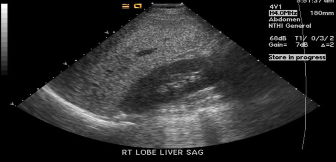
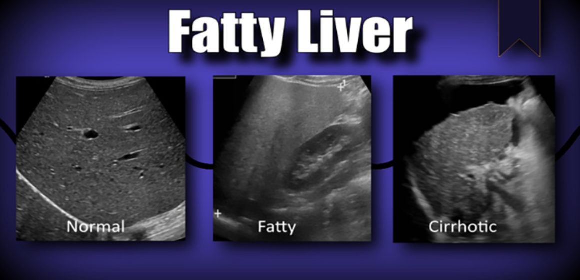
Objective: The purpose of this study was to categorize patients into different grades of nonalcoholic fatty liver disease (NAFLD) by ultrasonography and to compare the findings with their serum lipid profile. Materials and Methods: Descriptive, cross-sectional study design was used. One hundred and nine patients without a history of alcohol consumption of age more than 16 years attending general health checkup were selected at Tribhuvan University Teaching Hospital, Maharajganj, Kathmandu, as per the exclusion and inclusion criteria. Ultrasound scanning of the patients was done and their liver size, as well as grading of fatty liver, was done. Data were collected in predesigned pro forma and were analyzed using Statistical Package for the Social Sciences (SPSS) 16.0, IBM (SPSS Inc., Chicago, IL). Results: In this study, the mean age of fatty liver in males was found to be 44.3 years and in females was found to be 51.9 years. 22.9% of patients with NAFLD had increased liver size. Significant association with increasing grades of fatty liver was found with increasing levels of cholesterol (P = 0.028), low-density lipoprotein (LDL) (P = 0.017), liver size (P = 0.001), and body mass index (BMI) (P = 0.045) in patients diagnosed with NAFLD. No significant association with increasing grades of fatty liver was found with increasing levels of triglyceride (P = 0.32) and high-density lipoprotein (P = 0.25). Conclusion: Ultrasound is a safe and first-line modality for the evaluation of fatty liver and its grading. Increasing grades of fatty liver had significant association with increasing levels of cholesterol LDL, increasing liver size, and BMI of patients.
In summary, this study showed that fatty liver was more prevalent in male sex and age group of 40–50 years. Almost three quarters of fatty liver patients had normal liver size while one quarter had increased size of the liver. Almost half of the patients with fatty liver had increased BMI. Increasing grades
of fatty liver had significant association with increasing LDL, cholesterol, BMI, and liver size. No significant association of increasing grades of NAFLD was noted with increasing levels of serum TGL s or decreasing levels of HDL. Ultrasound is an important imaging tool for the diagnosis and grading of fatty liver. It is the first-line investigation modality for diagnosis of fatty liver. It is cheap, easy to use, and handle. However, it has some limitations like observer dependency. Next limitation of ultrasound in the cases of fatty liver is its inability to quantify the fat in the liver. Quantification of fat using CT scan or MRI may be more reliable for estimation and quantification of fat deposition in the liver. This can further be correlated with lipid profile in patients. Studies using CT and MRI for the quantification of fat and correlation with lipid profile can be done in the future. Findings in our study can be used for further management of patients with fatty liver. From the study, it was shown that increasing grades of fatty liver had significant association with deranged lipid profile. Deranged lipid profile is associated with cardiovascular problems. Hence, increasing grades of fatty liver has indirect relationship with cardiovascular problems.


